Saturday, December 4, 2010

FOOT DROP

Foot Drop


Introduction

Foot drop is a deceptively simple name for a potentially complex problem. Foot drop can be associated with a variety of conditions such as dorsiflexor injuries, peripheral nerve injuries, stroke, neuropathies, drug toxicities, or diabetes.
Common and superficial peroneal nerve, branches, ...

Common and superficial peroneal nerve, branches, and cutaneous innervation.


Deep peroneal nerve, branches, and cutaneous inne...

Deep peroneal nerve, branches, and cutaneous innervation.


The causes of foot drop may be divided into 3 general categories: neurologic, muscular, and anatomic. These causes may overlap. Treatment is variable and is directed at the specific cause.

History of the Procedure

Foot drop likely has been a problem for humans throughout our existence. Argument can be made that the biblical story of Jacob limping after wrestling with an angel in the book of Genesis represents the first recorded occurrence of foot drop.1

Problem

Foot drop can be defined as a significant weakness of ankle and toe dorsiflexion. The foot and ankle dorsiflexors include the tibialis anterior, extensor hallucis longus, and extensor digitorum longus. These muscles help the body clear the foot during swing phase and control plantar flexion of the foot on heel strike. Weakness in this group of muscles results in an equinovarus deformity. This is sometimes referred to as steppage gait, because the patient tends to walk with an exaggerated flexion of the hip and knee to prevent the toes from catching on the ground during swing phase. During gait, the force of heel strike exceeds body weight, and the direction of the ground reaction vector passes behind the ankle and knee center (see image below).
Diagram of the ground reaction vector during hee...

Diagram of the ground reaction vector during heel strike.


This causes the foot to plantar flex and, if uncontrolled, to slap the ground. Ordinarily, eccentric lengthening of the anterior tibialis, which controls plantar flexion, absorbs the shock of heel strike. Foot drop can result if there is injury to the dorsiflexors or to any point along the neural pathways that supply them.

Frequency

Peroneal neuropathy caused by compression at the fibular head is the most common compressive neuropathy in the lower extremity. Foot drop is its most notable symptom. All age groups are affected equally, but it is more common in males (male-to-female ratio, 2.8:1). Ninety percent of peroneal lesions are unilateral, and they can affect the right or left side with equal frequency.
A foot drop of particular concern to orthopedic surgeons is a peroneal nerve palsy seen after total knee arthroplastyor proximal tibial osteotomy. The estimated rates of this complication are 0.3-4% after total knee arthroplasty and 3-13% after proximal tibial osteotomy. Ischemia, mechanical irritation, traction, crush injury, and laceration can cause intraoperative injury to the peroneal nerve. Correction of a severe valgus or flexion deformity also has been suggested to stretch the peroneal nerve and lead to palsy. Postoperative causes of peroneal nerve palsy include hematoma or constrictive dressings.
In a study by Cohen et al, the relative risk of palsy was 2.8 times greater for patients who had received epidural anesthesia for total knee arthroplasty than for those who received general or spinal anesthesia.2 One postulation is that epidural anesthesia likely decreased proprioception and sensation, continuing to some extent postoperatively, allowing the limb to rest in an unprotected state susceptible to local compression. In addition, intraoperative neurologic damage may not have been readily apparent in the immediate postoperative period because of ongoing effects of epidural anesthesia. In this same study, the relative risk of palsy was 6.5 times greater in patients who had a prior lumbar laminectomy.
A series of patients who developed foot drop following primary hip arthroplasty were carefully examined and found to have spinal stenosis.3 Up to 70% of patients undergoing hip arthroplasty have electromyographic evidence of nerve injury, but they rarely have clinical symptoms.4 Patients with preexisting spinal stenosis are believed to be at increased risk for foot drop following hip arthroplasty because of this proximal compromise. This is the double-crush phenomenon described in more detail in the Pathophysiology section.

Etiology

Foot drop may follow direct injury to the dorsiflexors. A few cases of rupture of the tibialis anterior tendon leading to foot drop and suspicion of peroneal nerve palsy have been reported. This subcutaneous tendon rupture usually occurs after a minor trauma with the foot in plantar flexion.
Compartment syndromes also may lead to foot drop. These are surgical emergencies and are not associated only with fracture or acute trauma. March gangrene, a form of anterior compartment syndrome, is thought to be due to edema and small hemorrhages in the muscles of the anterior compartment occurring after strenuous activity in individuals not accustomed to it. Deep posterior compartment syndrome also may result in foot drop as a late sequela due to resultant contracture formation.
Neurologic causes of foot drop include mononeuropathies of the deep peroneal, common peroneal, or sciatic nerves. Lumbosacral plexopathy, lumbar radiculopathy, motor neuron disease, or parasagittal cortical or subcortical cerebral lesions also can manifest as foot drop. These lesions can be differentiated through clinical and electrodiagnostic examinations.
A common behavioral cause of foot drop is habitual crossing of the legs.5 These cases typically resolve with discontinuation of the habit.

Foot drop also may be seen as a combination of neurologic, muscular, and anatomic dysfunction. Charcot foot is one example.

Pathophysiology

The pathophysiology of nerve damage commonly causing foot drop is as follows:

  • The functional integrity of an axon and its target depend on the continued supply of trophic substances synthesized in the neuronal perikaryon and transported down the axon, known as axoplasmic flow.
  • A laceration interrupts this flow. A crush injury may compromise it as well.
  • A double-crush phenomenon occurs when a proximal insult in a nerve root diminishes axoplasmic flow, making it more susceptible to injury.
  • A distal lesion further compromises the flow, and clinical palsy results. This is the phenomenon thought to be responsible for the increased risk of foot drop after hip replacement in a patient with preexisting spinal stenosis. The spinal stenosis causes the proximal compromise, and intraoperative stretch of the sciatic nerve provides the distal insult.

Presentation

Direct injury to the dorsiflexors
With dorsiflexor injury due to laceration or contusion, both cause and effect are readily apparent on clinical examination. A young healthy or active healthy elderly patient usually benefits from surgical repair of the injury.
If the patient develops a degenerative rupture of the tibialis anterior muscle, foot drop may be observed, but the cause may not be immediately apparent. Such a patient is often an elderly man who suffers a minor trauma with the foot in plantar flexion. The patient stands with the foot everted and has some loss of dorsiflexion when attempting to heel-walk. The degree of foot drop varies depending on time elapsed since the rupture. Active function in the other muscles innervated by the deep and superficial branches of the peroneal nerve essentially rule out the possibility of a peripheral neuropathy. Functional recovery is achieved over time and is aided by bracing of the affected ankle. Surgery may not be required in this situation.

Compartment syndromes
Increased pain with passive stretch of the involved muscles is a consistent diagnostic indicator of a compartment syndrome. Pain out of proportion to the injury usually is the initial presenting symptom. Paresthesias follow, but at this point, irreversible myoneural injury has likely occurred. Foot drop also may be noted; the time of presentation varies with the compartment involved.
  • Anterior compartment syndrome
    • Clinical presentation of an acute anterior compartment syndrome includes pain with passive toe flexion, some weakness of toe extension, and diminished sensation in the first web space because of deep peroneal nerve compression.
    • The extensor hallucis longus usually is the first muscle to show weakness.
    • Anterior compartment syndrome may follow trauma to the extremity but also can be observed in march gangrene. Local erythema, heat, and brawny edema over the anterior compartment are present.
    • Regardless of the cause, wide fasciotomy of the anterior compartment must be performed to salvage the ischemic muscles.
  • Deep posterior compartment syndrome
    • An acute deep posterior compartment syndrome presents as pain and some weakness of toe flexion and ankle inversion. Pain on passive toe extension is referred to the calf.
    • Diminished sensation over the sole of the foot especially on the medial side is noted, resulting from posterior tibial nerve compression.
    • Foot drop develops because of ischemic contracture of the posterior compartment and is seen if the acute syndrome is not treated.
    • Once again, wide fasciotomy of the involved compartment is mandatory at the time of acute presentation.
  • Chronic compartment syndrome
    • This occurs in athletes in their third or fourth decade who have exercise-induced pain in the lower leg or foot within 20-30 minutes of beginning to exercise. Often, this occurs after a recent increase in intensity or duration of training or after a change in the training routine.
    • The symptoms resolve after 15-30 minutes of rest; however, as the syndrome progresses, pain occurs earlier and takes longer to resolve.
    • The anterior compartment is the most commonly involved.
    • Unless the patient has been exercising just before being examined, the physical examination may be nonspecific or normal.
    • Patients with a chronic anterior compartment syndrome may have diminished sensation in the first dorsal web space.
    • Recording of intracompartmental pressures before, during, and after exercise can provide useful diagnostic information as to which compartments may be involved. The following are believed to be indicative of the syndrome: a resting pressure of 15 mm Hg or more and/or a pressure of 30 mm Hg or more 1 minute post exercise and/or a pressure of 20 mm Hg or more 5 minutes post exercise. A slit catheter may be used to measure these pressures with the understanding that accuracy of the readings is influenced by depth of needle insertion; position of the leg, ankle, and foot; and force of muscle contraction.
    • Some preliminary investigation has been completed of MRI as a potential test for chronic compartment syndrome.
    • Nonsurgical treatment of a chronic compartment syndrome is only successful if the patient is willing to discontinue the inciting activity. The surgical treatment of choice is fasciotomy of the involved compartment.

Neurologic defects
Several neurologic defects can cause foot drop (see Introduction, Etiology). Equinovarus deformity associated with toe contracture is the most common lower extremity manifestation of stroke. This can be differentiated from a peripheral neuropathy on examination by eliciting hyperactive deep tendon reflexes and a positive Babinski sign. The patient's gait pattern can also suggest etiology. For example, patients with a paretic foot drop bear weight on the heel during initial foot strike, whereas those with a spastic deformity strike with the forefoot.
Another central nerve insult that can be associated with foot drop is L5 compression. In addition to weakness in the peroneal nerve distribution, the tibialis posterior is weak. Back pain, sciatica, and limitation of straight leg raising also are seen. Motor conduction velocity may remain normal.
Peroneal neuropathy also may be spontaneous, traumatic, or, less frequently, progressive. Peroneal neuropathy is characterized by weakness in dorsiflexion without back pain, sciatica, or other symptoms. Leprosy neuritis, for example, affects nerves where they are close to the skin and pass through a narrow fibrous or osseous canal. In addition to peroneal nerve palsy, patients with leprosy may have involvement of the posterior tibial nerve at the tarsal tunnel leading to anesthesia of the sole of the foot.

Foot drop from neuropathy may develop in patients who have undergone bariatric surgery, especially those who experience rapid postoperative weight loss.6 Micronutrient deficiencies (eg, vitamin B-12) may be a factor in these cases.7
Combination of neurologic, muscular, and anatomic dysfunction
These patients typically are diabetic and develop loss of protective sensation and proprioception, leading to unperceived trauma. This is coupled with an autonomic neuropathy that results in loss of sympathetic vasoconstriction and enhanced pedal blood flow, causing demineralization and subsequent bone weakness. Unperceived trauma, demineralization, and bone weakness culminate in destruction of the tarsal bones. This, in turn, forms a bony block at the ankle joint and foot drop. Progressive motor neuropathy is also present, in which the muscles weaken distally to proximally, resulting in loss of strength in the anterior compartment. The anterior muscles are overpowered by the Achilles tendon, leading to abnormal pronator stress at the midtarsal joint, further encouraging osseous breakdown and foot drop.


Relevant Anatomy

Fibers from the dorsal branches of the ventral rami of L4-S1 are found in the peroneal nerve, which is paired with the tibial nerve to constitute the sciatic nerve. The sciatic nerve leaves the pelvic cavity at the greater sciatic foramen, just inferior to the piriformis muscle.
The sciatic nerve bifurcates to form the peroneal and tibial nerves either at the distal third or mid-thigh level. The peroneal nerve crosses laterally to curve over the posterior rim of the fibular neck to the anterior compartment of the lower leg, dividing into superficial and deep branches. The superficial branch travels between the 2 heads of the peronei and continues down the lower leg to lie between the peroneal tendon and the lateral edge of the gastrocnemius. It then branches to the ankle anterolaterally to supply sensation to the dorsum of the foot (see image below).
Common and superficial peroneal nerve, branches, ...

Common and superficial peroneal nerve, branches, and cutaneous innervation.


The deep branch divides just after rounding the fibular neck. The initial branch supplies the anterior tibial muscle. Remaining branches supply the extensor digitorum longus and extensor hallucis longus and a small sensory patch at the first dorsal web space (see image below).
Deep peroneal nerve, branches, and cutaneous inne...

Deep peroneal nerve, branches, and cutaneous innervation.


The peroneal nerve is susceptible to injury all along its course. As part of the sciatic nerve, its funiculi are relatively isolated from those of the tibial nerve. Therefore, trauma to the sciatic nerve may only affect one of its divisions. Also, the funiculi of the peroneal nerve are larger and have less protective connective tissue, making the peroneal nerve more susceptible to trauma. In addition, the peroneal nerve has fewer autonomic fibers, so in any injury, motor and sensory fibers bear the brunt of the trauma. The peroneal nerve runs a more superficial course, especially at the fibular neck, also making it vulnerable to direct insult. It adheres closely to the periosteum of the proximal fibula, making it susceptible to injury during surgical procedures in this area
.

T.E.N.S

Transcutaneous Electrical Nerve Stimulation


Introduction


Transcutaneous electrical nerve stimulation (TENS) currently is one of the most commonly used forms of electroanalgesia. Hundreds of clinical reports exist concerning the use of TENS for various types of conditions, such as low back pain (LBP), myofascial and arthritic pain, sympathetically mediated pain, bladder incontinence,neurogenic pain, visceral pain, and postsurgical pain. Because many of these studies were uncontrolled, there has been ongoing debate about the degree to which TENS is more effective than placebo in reducing pain.
The currently proposed mechanisms by which TENS produces neuromodulation include the following:
  • Presynaptic inhibition in the dorsal horn of the spinal cord
  • Endogenous pain control (via endorphins, enkephalins, and dynorphins)5
  • Direct inhibition of an abnormally excited nerve
  • Restoration of afferent input
The results of laboratory studies suggest that electrical stimulation delivered by a TENS unit reduces pain through nociceptive inhibition at the presynaptic level in the dorsal horn, thus limiting its central transmission. The electrical stimuli on the skin preferentially activate low-threshold, myelinated nerve fibers. The afferent input from these fibers inhibits propagation of nociception carried in the small, unmyelinated C fibers by blocking transmission along these fibers to the target or T cells located in the substantia gelatinosa (laminae 2 and 3) of the dorsal horn.
Studies show marked increases in beta endorphin and met-enkephalin with low-frequency TENS, with demonstrated reversal of the antinociceptive effects by naloxone.6 These effects have been postulated to be mediated through micro-opioid receptors. Research indicates, however, that high-frequency TENS analgesia is not reversed by naloxone, implicating a naloxone-resistant, dynorphin-binding receptor. A sample of cerebral spinal fluid in those subjects demonstrated increased levels of dynorphin A.

The mechanism of the analgesia produced by TENS is explained by the gate-control theory proposed by Melzack and Wall in 1965.7 The gate usually is closed, inhibiting constant nociceptive transmission via C fibers from the periphery to the T cell. When painful peripheral stimulation occurs, however, the information carried by C fibers reaches the T cells and opens the gate, allowing pain transmission centrally to the thalamus and cortex, where it is interpreted as pain. The gate-control theory postulates a mechanism by which the gate is closed again, preventing further central transmission of the nociceptive information to the cortex. The proposed mechanism for closing the gate is inhibition of the C-fiber nociception by impulses in activated myelinated fibers.

Technical Considerations


A transcutaneous electrical nerve stimulation (TENS) unit consists of 1 or more electrical-signal generators, a battery, and a set of electrodes. The TENS unit is small and programmable, and the generators can deliver trains of stimuli with variable current strengths, pulse rates, and pulse widths. The preferred waveform is biphasic, to avoid the electrolytic and iontophoretic effects of a unidirectional current. The usual settings for the stimulus parameters used clinically are the following:
  • Amplitude - Current at a comfortable, low intensity level, just above threshold
  • Pulse width (duration) - 10-1000 microseconds
  • Pulse rate (frequency) - 80-100 impulses per second (Hz); 0.5-10 Hz when the stimulus intensity is set high
When TENS is used analgesically, patients are instructed to try different frequencies and intensities to find those that provide them with the best pain control. Optimal settings of stimulus parameters are subjective and are determined by trial and error. Electrode positioning is quite important. Usually, the electrodes are initially placed on the skin over the painful area, but other locations (eg, over cutaneous nerves, trigger points, acupuncture sites) may give comparable or even better pain relief.
The 3 options for the standard settings used in different therapeutic methods of TENS application include the following:
  • Conventional TENS has a high stimulation frequency (40-150 Hz) and low intensity, just above threshold, with the current set between 10-30 mA. The pulse duration is short (up to 50 microseconds). The onset of analgesia with this setup is virtually immediate. Pain relief lasts while the stimulus is turned on, but it usually abates when the stimulation stops. Patients customarily apply the electrodes and leave them in place all day, turning the stimulus on for approximately 30-minute intervals throughout the day. In individuals who respond well, analgesia persists for a variable time after the stimulation stops.
  • In acupuncturelike settings, the TENS unit delivers low frequency stimulus trains at 1-10 Hz, at a high stimulus intensity, close to the tolerance limit of the patient. Although this method sometimes may be more effective than conventional TENS, it is uncomfortable, and not many patients can tolerate it. This method often is considered for patients who do not respond to conventional TENS.
  • Pulsed (burst) TENS uses low-intensity stimuli firing in high-frequency bursts. The recurrent bursts discharge at 1-2 Hz, and the frequency of impulses within each burst is at 100 Hz. No particular advantage has been established for the pulsed method over the conventional TENS method.
Patient comfort is a very important determinant of compliance and, consequently, of the overall success of treatment. The intensity of the impulse is a function of pulse duration and amplitude. Greater pulse widths tend to be more painful. The acupuncturelike method is less tolerable, because the impulse intensity is higher.
The amount of output current depends on the combined impedance of the electrodes, skin, and tissues. With repetitive electrical stimuli applied to the same location on the skin, the skin impedance is reduced, which could result in greater current flow as stimulation continues. A constant current stimulator, therefore, is preferred in order to minimize sudden, uncontrolled fluctuations of current intensity related to changes in impedance. An electroconductive gel applied between the electrode and skin serves to minimize the skin impedance.

Medical complications arising from use of TENS are rare. However, skin irritation can occur in as many as 33% of patients, due, at least in part, to drying out of the electrode gel. Patients need to be instructed in the use and care of TENS equipment, with particular attention to the electrodes.
In some cases, individuals react to the tape used to secure the electrodes. Skin irritation is minimized by using disposable, self-adhesive electrodes and repositioning them slightly for repeated applications. The use of TENS is contraindicated in patients with a demand-type pacemaker, because the stimulus output of the TENS unit may drive or inhibit the pacemaker.
A variety of newer transcutaneous or percutaneous electrical stimulation modalities have emerged. They include the following:
  • Interferential current therapy (IFC) is based on summation of 2 alternating current signals of slightly different frequency. The resultant current consists of a cyclical modulation of amplitude, based on the difference in frequency between the 2 signals. When the signals are in phase, they summate to an amplitude sufficient to stimulate, but no stimulation occurs when they are out of phase. The beat frequency of IFC is equal to the difference in the frequencies of the 2 signals. For example, the beat frequency and, hence, the stimulation rate of a dual channel IFC unit with signals set at 4200 and 4100 Hz is 100 Hz.
  • IFC therapy can deliver higher currents than TENS can. IFC can use 2, 4, or 6 applicators, arranged in either the same plane, for use on such regions as the back, or in different planes in complex regions (eg, the shoulder).
  • Percutaneous electrical nerve stimulation (PENS) combines advantages of electroacupuncture and TENS. Rather than using surface electrodes, PENS uses acupuncturelike needle probes as electrodes, with these placed at dermatomal levels corresponding to local pathology. The main advantage of PENS over TENS is that it bypasses local skin resistance and delivers electrical stimuli at the precisely desired level in close proximity to the nerve endings located in soft tissue, muscle, or periosteum.3

Applications of Tens in Clinical Practice


Literature on the effectiveness of transcutaneous electrical nerve stimulation (TENS) in a variety of medical conditions reports a wide range of outcomes, from very positive to negative. Currently, there is an overall consensus favoring the use of TENS, with authorities differing on its value in different clinical situations. Generally, TENS provides initial relief of pain in 70-80% of patients, but the success rate decreases after a few months or longer to around 20-30%. To exclude a false-negative response, a trial of TENS for at least 1 hour should be given to confirm potential benefit from subsequent continuous use.
According to Johnson, the time from the start of stimulation to the onset of analgesia varies from almost immediate to hours (on average, 20-30 minutes in over 75% of patients and 1 hour in 95% of patients).8 The duration of analgesia also varies considerably, continuing only for the duration of stimulation in some patients and providing considerable, prolonged poststimulation relief in others. The same TENS protocol may have different degrees of antinociception in acute experimental pain compared with chronic clinical pain in patients with chronic low back pain (LBP).9
Patients differ in their stimulus preferences and in their rates of compliance. In Johnson's study of compliance in patients who benefited from TENS, 75% used the device on a daily basis. Patients showed individual preferences for particular pulse frequencies and patterns, and they consistently adjusted their stimulators to these settings in subsequent treatment sessions.
Indications for the use of TENS 
  • Neurogenic pain (eg, deafferentation pain, phantom pain), sympathetically mediated pain, postherpetic neuralgia, trigeminal neuralgia, atypical facial pain, brachial plexus avulsion, pain after spinal cord injury (SCI)
  • Musculoskeletal pain - Examples of specific diagnoses include joint pain from rheumatoid arthritis and osteoarthritis, acute postoperative pain (eg, postthoracotomy), and acute posttraumatic pain. After surgery, TENS is most effective for mild to moderate levels of pain, and it is ineffective for severe pain. The use of TENS in chronic LBP and myofascial pain is controversial, with placebo-controlled studies failing to show statistically significant beneficial results. A literature-study report from the American Academy of Neurology recommended against the use of TENS for the treatment of chronic LBP, stating that the strongest evidence indicates that it is ineffective against this condition Uncertainty also exists about the value of TENS in tension headache.
  • Visceral pain and dysmenorrhea - TENS has been successfully applied to these conditions as well
  • Diabetic neuropathy - A literature-study report from the American Academy of Neurology stated that TENS is probably an effective therapy for painful diabetic neuropathy and should be considered for use in the treatment of this disorder
  • Other disorders - TENS has been used successfully in patients with angina pectoris and urge incontinence, as well as in patients requiring dental anesthesia. Reports discuss the use of TENS to assist patients in regaining motor function following stroke, to control nausea in patients undergoing chemotherapy, as anopioid -sparing modality in postoperative recovery, and in postfracture pain.
Contraindications for the use of TENS 
  • TENS should not be used in patients with a pacemaker (especially of the demand type).
  • TENS should not be used during pregnancy, because it may induce premature labor.
  • TENS should not be applied over the carotid sinuses due to the risk of acute hypotension through a vasovagal reflex.
  • TENS should not be placed over the anterior neck, because laryngospasm due to laryngeal muscle contraction may occur.
  • The electrodes should not be placed in an area of sensory impairment (eg, in cases of nerve lesions, neuropathies), where the possibility of burns exists.
  • A TENS unit should be used cautiously in patients with a spinal cord stimulator or an intrathecal pump.

Comparison Between Tens and Other Electrical Modalities


A number of studies have compared transcutaneous electrical nerve stimulation (TENS) with similar therapeutic modalities, including percutaneous electrical nerve stimulation (PENS), interferential current therapy (IFC), and acupuncture. The results included the following:
  • In one study of elderly patients with chronic low back pain (LBP), acupuncture and TENS had demonstrable benefits, with the acupuncture group demonstrating improvement in spinal flexion.
  • In patients with chronic LBP and sciatica, PENS was more effective than TENS in providing short-term pain relief and improved function, including an improved quality of sleep and sense of well-being.
  • Overall, 91% and 73% of patients, respectively, chose PENS as the preferred modality for pain relief in LBP and sciatica.
  • PENS has been used successfully for pain relief in patients with acute herpes zoster and in persons suffering from cancer with bony metastases.
  • IFC and TENS had a statistically significant effect on the median nerve excitation threshold in young women

    .

LOW BACK PAIN

LOW BACK PAIN


DEFINITION

            Pain on the lumbar area caused by an underlying disease. It is not a disease but a symptom. Pain between the costal margins and gluteal folds.          
      

Anatomy

·         The kidney shaped lumbar vertebral body is wider laterally than it is deep anteroposteriorly, and broader than it is high. Its periphery is deeply hollowed out in a shape of a diabolo, except posteriorly where it is nearly flat.
·         The two (2) laminae are highly set and run posteriorly and medially but they lie in a plane which is oblique inferiorly and laterally.
·         The transverse processes, better called ostiod process since they are in fact rib vestiges.
·         They are attached at the level of the articular processes and run an oblique course posteriorly and laterally.
·         The pedicle, a short and bony segment joining the vertebral body to the vertebral arch and attached to the former at its superolateral angle.
·         The superior articular process lies on the superior border of the vertebral arch near the junction of the lamina with the spinous process.
·         The vertebral body of L5 is higher anteriorly than posteriorly so that its profile is wedge-shaped or even trapezoidal with its longer side lying anteriorly.

Spine
·         An aggregate of superimposed segments that can be termed functional units.

Functional Unit:
·         Composed of two vertebral bodies, one superincumbent on the other, separated by the IV disk.

Anterior weight bearing portion of the functional unit:
·         Comprised of two adjacent rounded vertebral flattened at their cephalad and caudal ends.
·         At birth and throughout maturation, caudal and cephalic ends of the vertebra are slightly convex and coated with cartilage, undergoes ossification from age 15-20 and fuse with the vertebral bodies.
·         Ossification layer at each endplate called epiphysis, remains active throughout growth; influence by Wolff’s law

Intervertebral disk
·         80% of load
·         Hydrodynamic elastic structure that is imposed between two adjacent vertebrae; shock absorbing mechanism
·         25% height of the spine
·         90% composed of water- decrease with age
·         65% water in old age

·         Central (nucleus) pulposus:
o   Ovoid, yellowish and gelatinous in appearance
o   Middle part of the IV disk
o   Made of mucoprotein
o   Proteoglygcans- responsible for water retention
§  Greater than collagen

·         Annulus fibrosus
o   20 firm, concentric rings of collagen surrounding the nucleus pulposus
o   3 layers of fibrocartilage:
§  Sharpey’s fiber- outermost layer
o       Collagen > proteoglycans
·          Nucleus- homogenous, elastic globoid centrally located within a fibrocartilagenous envelope most disk hydration occurs via imbibition.
·         The disk in early human development is nourished by direct blood supply; upon maturation, nutrition is by primarily imbibitions, and to a lesser extent, osmosis.
·         Annular fibers fail after exceeding 5 degrees of rotation, site of failure varies because of the differences in angulation where the inner fibers have more vertical orientation.

Posterior portion of the functional unit:
·         20% of load
·         Forms the outer wall of the spinal canal
·         Neural arch extends posteriorly as pedicles to form as lateral transverse process
·         Widens to form the zygaphophyseal(facet) joint (note that the lumbar zygapophysis lie I the sagittal plane, which allows flexion and extension, markedly restricts rotation and lateral flexion)
·         Laminae continues postermedially to rejoin centrally and form the posterior superior spine
40-50% of the torque strength of the whole joint is providedby the disk annulus, the rest is provided by the posterior articulations: (the facets, their capsules and interspinous ligament)

*as the lumbar spine laterally flexes, there is mandatory, simultaneous rotation toward the side that the spine bends:
1. facets of the concave side approximate
2. facets on the side from which the spine is bending separate
3. IV disk is sheared laterally with a degree of rotation
4. normal movement does not change the width or the content of the foraminal to an abnormal unphysiologic degree

Ligaments of the Lumbar Spine
In the sagittal section there are two (2) sets of ligaments:
·         The anterior longitudinal ligament- stretches as a long dense band from the base of the occiput to the sacrum on the anterior aspect of the vertebrae.
·         The posterior longitudinal ligament- stretches from the base of the occiput to the sacral canal. It tapers at the level of L2 ( reason why posterolateral herniation is more common)
Segmental ligaments that joins the vertebral arches:
·         Ligament Flavum- inserted inferiorly into the superior border of the underlying lamina and superiorly into the medial aspect of the overlying lamina.
·         Interspinous ligament
·         Supraspinous ligament
·         Intertransverse ligament

Muscles
    Erector Spinae (sacrospinalis)
Origin: last 2 thoracic vertebrae, lumbar vertebrae, sacral spine, sacrum, SI ligament, medial aspect of the iliac crest.
            Spilts into three(3) columns:
·      Iliocostalis (lateral)- inserts into the angle of the rib cage (C4-T6)
·      Longissimus- inserts in the transverse processes cephalad to T1 (only one to reach the skull)
·      Spinalis(medial)- flat, aponeurosis muscle. Insertion at the posterior spine of the thoracic vertebrae

     Transverse Spinae (underneath the erector spinae)
            1. Semispinalis ( superficial)- origin: tips of the transverse processes
                                                           Insertion: tips of the posterior spinous process; Spans 3-5 segments
2. Multifundus- between the spinous and transverses crest, from overlying erector spinae aponeurosis           and from all the transverse processes up to C4 vertebra
-spans 3 vertebral segment- attach to the superior border of the next transverse process above it.
            3. Rotatores- spans only one segment; origin: transverse process of one vertebrae to the spinous  process of the adjacent vertebrae.


            Other muscles:
            1. interspinalis- between two adjacent spinous process
            2. intertransversii- between adjacent transverse process

Thoracolumbar fascia
·         deep investing membrane which covers the deep muscles of the back of the trunk. It is made up of three layers, anterior, middle, and posterior. The anterior layer is the thinnest and the posterior layer is the thickest. Two spaces are formed between these three layers of the fascia. Between the anterior and middle layer lies the quadratus lumborum muscle. The erector spinae muscle is enclosed between the middle and posterior layers.


Angles of the Lumbar Vertebrae:
1. Angle of the sacrum- approximately 30 degrees
                                    - measured from the horizontal line toward the superior surface of S1.
2. Angle of Pelvic Tilt- approximately 60 degrees
- from the horizontal line across the line within the promontory of sacrum and symphisis.
3. Lumosacral angle- approximately 30 degrees
                                - angle from the axis of L5 to S1


Lumbar pelvic Rhythm
·         It is the coordinated movement of the lumbar spine and pelvis
·         The lumbar spine has 5 functional units, each unit flexes approximately 9 degrees (5 units= 45degrees), allowing the person to bend over forward only a portion of the amount of flexion required for toe touching.
·         For additional forward flexion, a simultaneous rotation of the pelvis must occur.
·         Sequence of activities occurring within the functional units:
a. during flexion:
           1. extensor muscle relaxed and 3 physiologic curves becomes straight
           2. upon flexion head moves ahead of the COG, there is contraction of the erector spinae muscles
           3. further forward flexion, lumbosacral spine goes to kyphosis.
           4. extensor muscles elongates smoothly and gradually.
           5. erector spinae muscles goes eccentric contraction, each functional unit flexes about 9 degrees
           6. hip extensors maintain isometric contraction
           7. when back muscles and ligament become taut, full lumbar khyphosis has been reached.
           8. pelvis begin forward rotation by elongation and deceleration of gluteus maximus and hamstrings to give further way to flexion
           9. full flexion has been achieved, the pelvis is stricted by its ligaments and facial tissue.
           10. no muscular activity, only fascia and ligaments are operative.
b. during re-extension:
           1. pelvis should rotate first before the lumbar spine resumes its lordosis.
           2. the hips and the knees should be slightly flexed.
           3. resumption of the erect posture without excessive rotation and de-rotation.


Pain Free structures:
1. IV disk (nucleus pulposus, inner and middle layer of annulus fibrosus)
2. Vertebral body
3. nerve root (per se)- irritation will only cause paresthesia, dysthesia, paresis but rarely pain
4. Ligamentum Flavum

Pain sensitive structures:
1. outer layers of annulus fibrosus
2. Anterior Longitudinal Ligament
3. Posterior Longitudinal Ligament
4. Recurrent nerve from nerve root
5. Erector Spinae
6. Other posterior supporting ligaments
7. Zygapophyseal joints

Disc Pressure in various postures:
Supine                                                           25%
Side lying                                          75%
Standing Upright                              100%
Sitting upright                                               140%
Standing while bending forward   160%
Sitting while bending forward                    185%
Standing while bending forward
   and holding weights                                 220%
Sitting while bending forward
   and holding weights                                 275%


Types of Low Back Pain:

1. Static LBP
·         Excessive lordosis or “sway back” posture has been considered and is responsible for 75% of all static or postural LBP
·         Pain is produced by:
o   Approximation of the facets with compression
o   Intervertebral foramen closes and encroaches the nerve root dura
o   Disk can bulge posterolaterally, putting the strain on the posterior longitudinal ligament
·         When approximation of the post articulation causes sliding of the facets (kissing of the spine) into each other, they become weight bearing which is not its physiologic function, resulting in synovial inflammation leading to pain.
·         Excessive lordosis is usually seen in:
o   Pregnant women who stand with arched back
o   Improper sitting position
o   Persons wearing high heeled shoes
o   People with poor sleeping habits (prone positioning)
o   Occupation requiring hours of sitting and standing (i.e delivery truck drivers, people who do ironing)

2. Kinetic Low Back Pain
·         Implies irritation of pain- sensitive tissues activated by movement of the spine
·         Caused by improper functional use of lumbosacral spine
·         Impaired lumbar pelvic rhythm
·         Pain can originate in these manners:
o   Normal stress on an unprepared back
§  Lack of anticipation and preparation causing contradiction to “overshoot the mark”
§  Excessive movement exceeds the physiologic limitation imposed by the ligaments
§  Micro or macocapsular and articular tissue damage occurs resulting in pain and disability
o   Abnormal stress on a normal low back
§  By extraordinary weight imposed on the supporting structure
§  By average weight held in a markedly eccentric manner
§  By light object held for unduly lone period of time
ü  Forward flexed standing or sitting posture of 10 -15 degrees
§  Cause of industrial cases of LBP
o   Normal stress on an abnormal low back
§  Conditions such as structural scoliosis, tight hamstrings, and osteoarthritis


EPIDEMIOLOGY

ü  Second to headache as leading cause of pain
ü  Leading cause of expense in workers compensation
ü  60-90% lifetime rate
ü  >5 % annual rate
ü  Men=women
ü  >1% with lumbar radiculopathy
ü  Age of lumbar surgery=45
ü  5th highest reason of hospitalization
ü  Most episodes resolve with or without treatment


ETIOLOGY

A. Congenital disorders
o           Sacralization of lumbar vertebrae
o           Lumbarization of sacral vertebrae
o           Tropism
§         Assymetrical segmental scoliosis usually in the lower lumbar segments
§         Nerve root on the side of concavity prone to entrapment
§         L4-L5, L5-S1
§         Back area may present with tenderness upon palpation
§         Radicular pain
§         Paresthesia if with nerve root entrapment
§         Agrrevated by extension with lateral flexion to the concave side
§         Relieve with flexion and lateral flexion towards convexity


B. Tumors
o          Benign
§        Involving nerve roots of meninges
§        Involving the vertebrae
o          Malignant
§        Primary malignant tumors (multiple myeloma-most common)
§        Secondary malignant tumor (metastatic- most common tumor)
-                                    Metastatic cancer from the lungs, prostate, breast, kidneys, and colon.
-                                    Symptoms: pain especially at night
Treatment:
·                             Radiation therapy
·                             Decompression bracing
·                             Ambulatory assistive device
C.  Trauma
o Lumbar strain
o Fractures
o Spondylolysis- fracture of the pars interarticularis
-                                                 Unilateral
-                                                 Commonly involving L5 and L4
-                                                 Male> female
Etiology:
§                   Stress Fracture
§                   Congenital failure of fusion during maturation of the bones
§                   Birth fractures
§                   Increase lumbar lordosis
§                   Impingement of the articular process on the pars interarticularis
§                   Weakness of the supporting muscles
§                   Pathologic changes in the pars interarticularis
§                   Axial loading of only about 600 lbs is sufficient to produce fracture in the isthmus of the neural arch if the spine is maintained in hyper lordosis

Pathophysiology:
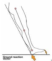
§   The gravitational and muscular loads acting across the step in inclined of the upper surface of the sacrum can be resolved into:
·                                                               Shearing components( L5 displaced forward on S1.
·                                                               Compressive component
ü  at right angles
ü  in accordance with Newtons 3rd law
- The mutual forces of action and reaction between two bodies are equal, opposite and collinear. This means that whenever a first body exerts a force F on a second body, the second body exerts a force −F on the first body. F and −F are equal in magnitude and opposite in direction. This law is sometimes referred to as the action-reaction law, with F called the "action" and −F the "reaction".

Signs and symptoms:
§               localized lumbosacral pain that is usually worst with extension and partially relieved with flexion
§               local tenderness
§               spasm


Diagnosis
§               Xray- degenerative and hypertrophic changes can be found
§               CT scan
§               MRI

Treatment
§               Rest
§               Rigid mobilization(e.g. Modified Boston overlap brace)- worn 23 hrs per day for 6 mos in cases of symptomatic spondylolysis

o  Spondylolisthesis- forward or backward shearing subluxation of the body of a superior vertebrae on its caudal counterpart
-    Most common L5 on S1, and L4 over L5
-    Grade according to displacement:
I        forward displacement of </= to 25%
II       forward displacement of 26-50%
III      forward displacement of 51-75%
IV     forward displacement of >75%
Etiology:
§ 5th lumbar vertebrae tends to glide forward due to shear force and gravity
§ Forward sliding is normally prevented by mechanical alignment and relationship of posterior facet joints, their ligaments, capsule, integrity of IV disk and of the longitudinal ligaments.
·      Intact nucleus pulposus expand the disk and places the annulus fibers and longitudinal ligament under tension that resist vertebral sliding
·      The contact of the facets and their planes also resist anterior subluxation of the vertebrae

§ Defect in any of the supporting structures may permit listhesis of the superior vertebrae over the inferior vertebrae of the involved functional unit.
ü    5 categories according to causative factors:
1. Isthmic-  most common; occurs due to spondylolysis of the pars interarticularis
2. Dysplastic or congenital- caused by dysplasia of the facet joint of the upper sacrum, leading to inability to resist shear stresses, then forward slippage occurs
3. Degenerative- seen in older spine, related to long standing intersegmental instability from degenerative facet or disk disease; most common in L4 and L5 level.
4. Traumatic- rare; cause by acute fracture secondary to trauma
5. Pathologic- secondary to metabolic and malignant or infectious disease.

§ Other causes:
·      Backward bending(e.g. Gymnastics)
·      Obesity and disk degeneration
·      Congenital asymmetrical facets

Clinical Manifestations:
§ LBP with referred pain laterally to the region of the SI joints
§ Pain radiate to hips, thighs, and even to the feet, not precisely in dermatomal pattern
§ Stiffness with limited flexibility
§  Presence of palpable “ledge” on LS spine
§ Segmental Lordosis


Diagnosis
§ Xray and CT scan may reveal:
·   Defect in pars interarticularis
·   Elongated arch
·   Fracture
·   Severe LS angulation
·   Severe IV disk degeneration

§ Lateral view of LS spine reveals degree of subluxation
·   Foraminae are deformed and sciatic radiculopathy may result
·   Normal foraminae narrows during the process of extending the lumbar spine
·   Narrowing is enhanced and nerve entrapment is more probable

Treatment
§ Surgery- when there is neurologic deficit
·         Anterior interbody fusion between L4-L5 or L5-S1
·         Laminectomy- if neural arch is involved

§ PT management
·                                                           Pelvic tilting
-                                                            Exercise in which ant. aspect of the pelvis is elevated and the posterior lowered
-                                                            Strong abdominals, especially the obliques must be gained and maintained
-                                                            Avoid excessive lordosis
-                                                            Extensor muscle must be strengthened
·                                                          Corset or braces
-                                                           Decreases lordosis
-                                                           Reinforces the abdominal component
-                                                           Minimizes excessive motion during lifting and bending
-                                                           Must be used as an adjunct to exercise not to replace it.

D.  Metabolic Disorder
o Osteoporosis- decrease bone mass per unit volume of bone
o Osteomalacia- disturbance in calcium metabolism

E. Inflammatory Diseases
o Rheumatoid arthritis
o Ankylosing spondylitis (bamboo spine)
Ø  Marie strumpell- ascending
Ø  Bechterew- descending

F. Degenerative Disorder
o                      Spondylosis- kissing of spine
§ Degenerative or osteoarthritic changes in the spine
§ Stiffness is the main feature
§ Dull and nagging pain and stiffness in the morning
§ Treated by resting
§ Induced by lumbar extension

Treatment:
§ avoid hyperextension
§ weight control
§ isometric exercise of the quadratus lumborum and abdominals
§ avoid sleeping prone
§ LS support for severe pain( avoid use >3 weeks)
§ NSAIDS
§ Facet joints injection

o Osteoarthritis- wear and tear
o Herniated disk- disk protrudes backward and compresses the nerves that are distributed to the LE.
- 90% occurs in the region of L4-L5, L5-S1 disk spaces
- Found I patients between 30-50 y.o
Types:
a. Protrusion
-                                                      Most common
-                                                      Involves no tearing of the annulus fibrosus with some bulging of the nucleus pulposus

b. Prolapse
-       Complete tear through the annulus fibrosus but with an intact anterior &/or posterior longitudinal ligament
-                                                      Only the outermost fibers of the annulus contains nucleus

c. Extrusion
-       Complete tear through the annulus fibrosus with involvement of the longitudinal ligaments
-                                                      Annulus fibrosus is perforated and discal materials moves into the epidural space

d. Sequestration
-       Formation of discal fragment from the annulus fibrosus and nucleus pulposus outside the disk proper.

Etiology:
§ Herniation of the nuclear materials usually occurs after the onset of degenerative changes
§ Most rupture occur in the 3rd of 4th decade
§ Rupture result of combination of acute trauma and pre-existing degenerative changes
§ Poor conditioning and faulty biomechanics

Clinical Manifestations:
§ Recurrent episodes of back pain and pain in the lower leg
§ Prolonged sitting or standing can aggrevate the pain
§ (+) sciatic pain/ radicular pain
§ Increase in intra spinal pressure caused by coughing, sneezing, or bending down and during bowel movements
§ Spinal movement also aggrevate the pain

Treatment
§             Surgical
·   Laminectomy
·   Laminotomy- division of a lamina of a vertebrae
·   Nucleotomy

§             Pt management
·   Icing for 15 mins (acute onset)
·   Heating modalities after 24-48 hrs.
·   Intermittent lumbar traction of nerve root pain( pain radiating down the legs)
·   PRES on abdominals
·   Postural control
·   Use of proper body mechanics
·   Use of a plaster jacket to provide rest and security to the spine for 3-4 weeks

o Spinal Stenosis- localized narrowing of the spinal canal from the structural abnormality of each bony components
-                                                      Canal may be narrowed by reduction of its transverse diameter, AP diameter, or both.
-                                                      Narrowing of the central portion of the spinal canal gives rise to central stenosis and the narrowing of the lateral part of the nerve canal.
-                                                      Give rise to lateral stenosis with lateral body entrapment
-                                                      Greatest central canal narrowing occurs at L3-L4 and L4-L5 regions.

            Etiology:
§ Congenital- primary
Ø                               Idiopathic
Ø                               Achondroplastic- pedicles are short and the interpediculate space is decreased.
§ Acquired- secondary
Ø       Spondylolisthetic- pars interarticularis defect secondary to DJD
Ø  Iatrogenic- post diskectomy, post fusion
-          Metabolic disease like Paget’s Disease(chronic skeletal disease characterized by progressive structural changes and typical deformities occurring in long bones, spine, pelvis, and cranium. 

            Pathomechanics
§ Narrowing of the lumbar spinal canal
§ Degenerative changes can lead to further narrowing
§ Lumbar spine becomes stenotic
§ (+) leg pain when walking

            Clinical Manifestations:
§ Pseudoclaudication( neurologic claudication)
-    Paresthesias of one or both legs occurring after a period of distance walking or after a period of standing.
-    Symptoms subsides then disappear after sitting
-    As the symptoms gradually progress, weakness of the legs may interfere with normal gait

                              Factors that lead to pseudoclaudication:
a. Exercise of the legs normally causes an increase venous capacity, dilatation of veins that accompany the nerve roots as they emerge to the foraminal gutters
b. If there is compression of nerves within the foramina, there is arterial supply limitation compromised of the venous return.
§ Sitting flexed forward causes a lumbar kyphosis and the following results:
-    canal lengthens
-    cauda equina fibers elongates
-    facets separate and the foraminae opens
-    Venous capacity and blood flow to the nerve returns

            Diagnostic procedures:
§ CT scan
                              -Most effective in identifying spinal stenosis
§ EMG
                              -Provide additional evidence of nerve root involvement
§ Xray findings:
·                                                                                                       Lateral view: shortening of the lower lumbar pedicles
·                                                                                                       AP view: reduce distance between pedicles

            Differential Diagnosis

INTERMITTENT CLAUDICATION
PSEUDOCLAUDICATION
Pain on calves (distal to proximal)
Pain on lumbar spine – calf
Elicited by walking lone distance & exercise
Elicited after prolonged standing
Pain is usually felt as squeezing and cramping
Felt as numbness or burning; rarely cramping
Relieved by cessation of activities
Relieved by sitting and stooping forward

            Treatment
§ Medical management            
Ø  Oral steroids and epidural steroids
§ Surgical management
Ø Laminectomy- surgical removal of the lamina
Ø Foraminectomy- surgical removal of the foramen
§ PT Management
§ Williams Exercise- abdominal strengthening
§ Lumbosacral support- ex. Corset
§ Pelvic and Gravity Traction
§ Rest
G. Mechanical Causes
o                              Intrinsic factors
§ Poor muscle tone
§ Myofascial pain
§ Instable vertebrae
§ Hyperlordosis

o Extrinsic factors
§ Renal problem
§ Hip problem
§ Scoliosis

 DIAGNOSIS

A.   Clinical

1. History
a.  mode of onset
b.  aggrevating and relieving factors
c.  effect of posture, exertion, rest
d.  effect of cough, sneeze, strain
e.  presence or absence of pain at night
f.   course of pain
g.  history of back or lower limb pains
h.  associated lower limb symptoms
i.    urinary frequency, urgency, retention, bowel/bladder incontinence
j.    history of lumbar surgery
k.  treatment implemented, medications used, and effects of these medications
l.    presence or absence of litigation or compensation issues

            2. Examination

·   Inspection
a.  Deformities
b.  Paraspinal spasms
c.  Birthmarks
d.  Unusual hair growth
e.  Listing to one side
f.   Decrease or increase lordosis
g.  Scoliosis
h.  Atrophy
i.    Asymmetries

·   Palpation and percussion
a.  Trigger points
b.  Local tenderness
c.  Spasm
d.  Tightness
e.  Withdrawal to touch

·   ROM of lumbar spine
a.                   Flexion- 40 degrees
b.                   Extension-15degrees
c.                   Lateral bending- 30 degrees
d.                   Rotation-40 degrees

·         Neurologic Exam
a.    Gait and coordination
b.    Muscle stretch reflex
c.    Muscle bulk
d.    Muscle strength
e.    Sensory examination
            3. Special tests

1.        Neurologic Dysfunction
a.          SLR/Lasegue test
-    Passive flexion of the hip until pain is elicited
-    0-30 degrees- muscular
-    30-70- neuropathic
-    >70 degrees- skeletal

 b. Brunzinki’s Sign/ Hyndman’s/ Lindner’s/Soto-hall sign
-          Flex neck until the chin is on the chest


c. Braggard’s test
-          Foot DF
-          Pain with neck flexion and foot DF- stretching the dura mater
-          Foot DF alone- hamstring tightness

d. Sicard’s test
-          SLR and extension of the big toe

e. Turyn’s test
-          Extension of the big toe

f. Bilateral SLR
-          Pain <70degrees- SI joint lesion
-          Pain >70 degrees- Lumbar spine lesion

g. Well leg raising/ Lhermitte’s / Cross over sign
-          Space occupying lesion- herniated disk

h. Prone knee bending/ Nachlas test
-          Passively flex knee while in prone
-          Unilateral lumbar pain- L2-L3 nerve lesion
-          Anterior thigh pain- quadriceps tightness
-          SI/ lumbar pain- tight rectus femoris

i. Kernig- Brudzinski’s Test
-          Flex head unto chest while supine
-          Flex hip with legs extended until pain is felt
-          Flex knee until pain disappear
-          Significance- meningeal, nerve root or dural irritation

      2. Test for Spinal Theca Compression
                                    a. Naffzigger’s Test
                                    b. Valsalva’s maneuver
                                    c. Milgram’s test

                        3. Test for SI pathology
                                    a. Patrick/FABER Test
                                    b. Gaelen’s Test
                                    c. Pelvic Rock Test

                        4. Test for muscle tightness
                                    a. Thomas Test
                                    b. Ober’s Test

B.   Diagnostic Testing

Ø Plain Radiography
·         Fracture
·         Dislocation
·         Spondylolisthesis
·         Degenerative disease
·         Narrowing of IV disk spaces
·         Tumors of he spine


Ø Radiostope Bone Scaning
·         Tumors( bone metastasis)

Ø CT scan and MRI
·         Disk disease
·         Herniated or extruded disk
·         Tumors
·         CT scan for bony lesion
·         MRI for epidural, intradural, intraaxial spinal cord lesion

Ø MRI Electromyography
·         Neurogenic changes and denervation
·         Provides physiologic exam
           
DIFFERENTIAL DIAGNOSIS

Rickets

            In the presence of a spinal curve that was distinguished by the alteration of the shape in the other bones with which it is accompanied, especially in those in the lower limbs and the chest one could make the diagnosed rickets of rachi is, rickets a wide spread disorder, was sometime referred to as the “depressed of the spine.

Stooped Posture
         
             A” hooped like curvature” could occur in rheumatism, however, a smoothly rounded anterior curvature could also be congenital or developmental. Some people, either  from their original conformation or from early habit of stooping, present the appearance of a very considerable gradual or hoop-like curvature.

Tuberculosis

            It was especially important to differentiate rheumatism, rickets, and more postural deformities form bony tuberculosis, an infection frequently encountered in those days. Tuberculosis, referred to as scrofula or white swelling, was identified in the spine on the basis of a kyphosis that instead of being smoothly rounded, was interrupted by, an angle projecting posteriorly. Such an angular curvature can be produced into other way, and can never be mistaken.

Syphilis

            The other infection disease one had to keep in mind was syphilis, although not as common cause of back pain. It was usually encountered in the skull, tibia or ulna. Pain cases showing skeletal deformities, especially those presenting with a deformed and painful shin bone, were diagnosed as vascular musculo rheumatism resulting in either from a syphilic cause or from the aggressive treatment of syphilis with mercurial drugs, in which case it was known as mercurial rheumatism.

Scoliosis

            Known as lateral curvature of the spine, scoliosis was often encountered especially in girls. The direction of the curve made the diagnosis easy.


MEDICAL MANAGEMENT

            Medications should be taken 4-6 hours. Taking the medication whenever necessary can cause drug dependency.
·         anti-inflammatory drug blocks the transmission of substances that could imitate soft of                                           the low back.
·         NSAIDS provide anti-prostaglandin B formation
·         steroids for severe pain
·         muscle relaxant muscular tension compresses all the inflammed tissues and restricts movements which cause pain but is questionable since muscle spasm is protective in nature
·         sedative tranquilizer
·         anti-depressant endorphin formation
·         intramuscular injections and injection of an anesthetic agent with soluble steroid injected into the multifidus triangle which frequently interrupts the painful low back pain

SURGICAL MANAGEMENT

Surgical approach and techniques are indicated assuming that:
·         patient has been correctly selected  as suitable operation
·         proper conservative treatments has failed
           
Approaches can be subdivided into:
Anterior Trans operational app.
·   used for 1.5 SI fusion
·   also used osteomyelitis and tumors involving L5-S1
·   can't be used in decompressing the neural elements

Anterior Intra operational app.
·   provides a wide exposure
·   all operation involving the vertebral body and disc from L2-S1
·   better for L4-5 and L5-S1 fusion
·   advantage iliac grafts can be taken through the same skin incision
·   posterior midline app.
·   all posterior elements, transverse processes, spinal canal, disc spaces and vertebral bodies and be exposed for surgical techniques enumerated below.

To operate merely to relieve pain is not and should not be an indication for surgery. Fear in the mind that pain is very subjective

1) Laminectomy and/or Laminetomy
- is the surgical approach to seeing and ultimtely removing the osteophytes
- laminetomy implies removing asufficient portion of the lamina to view the nerve within    the neural    canal or within the foramen
- laminectomy implies removing all the half of the lamina thus giving the target view of the nerves  of wider in the neural canal to free the nerve roots
2) Foraminectomy
- is a technique of widening of foramena that has been confirm to be narrow or deform bone thus being too narrow for the emerging nerve root


PHYSICAL  THERAPY MANAGEMENT

Goals of final rehabilitation

·         Full, pain free ROM of the injured and adjacent ligaments as well as hip, girdle, spinal and lower extremity structures that influence the lumbar spine.

·         Optimal strength, endurance and coordination of the neuromuscular system affecting the lumbar  spine.
·         Prevention of further injury and recurrence
·         Return to normal functional activities

Acute phase goal
§  decrease pain and inflammation
§  protect injured area from further injury

Subacute phase goals
§  regain soft tissue flexibility and segment motion
§  restore full function to injured and supporting structures
§  maximize lower extremity muscular flexibility for normal lumbar motion
§  optimal joint mobility

Chronic phase goals
§  multidisciplinary pain management
§  improve functional ADL's
§  discontinue use of health care system
§  pain control


Treatment Outline

Acute (0-4 weeks);
§  rule out serious problems
§  short course of bedrest
§  posture connection exercise
§  lumbar support
§  decrease painful/inflammation/spasm
§  keep them active
§  some pain is okay
§  teach patient what to avoid


Subacute (1-10 weeks)
§  increase mobility
§  strength, flexibility, and fitness
§  body mechanics training
§  back care education
§  work stimulation
§  posture training
§  evaluate work environment
§  return to work

Acute/ Subacute pain
§  cold packs, hot packs
§  bed rest
§  simple analgesics
§  massage
§  TENS/US
§  Corset/brace
§  Pelvic traction
§  Manipulation
§  Early activity
§  Back exercises

Chronic (after 7-10 weeks)
§  increase work simulation
§  increase strength, flexibility, fitness
§  rehabilitation psychology
§  behavior modification

William’s Flexion Exercise
1. pelvic tilt
2. single and double knee to chest
3. partial and full sit ups
4. Straight leg raises
5. hurdle’s position
6. wall slides

McKenzie Exercise
1. side gliding
2. forward bending
3. passive prone extension
            *on elbows
            *on hands
4. Knees to chest while supine
5. prone lateral shifting of the hips off midline
6. backward bending
7. flexion while sitting
8. slouch-over correct

Low Back Hygiene
1. During prolonged sitting
·         get up every 2o minutes
·         perform pelvic tilt exercise
·         sit with knees bent
·         one or both feet slightly elevated

2. Driving
·         bring seat close to steering wheel
·         knees slightly higher than the hip
·         stretch every 20-30 minutes

3. Before coughing or sneezing
·         tighten abdominal muscles

4. Begin progressive low back isometrics and stretching

5. Bend knees when picking an object from the floor

6. When getting into bed, sit on the edge of the bed, turn and roll to one hip, bring knees up and slowly recline.

Prevention of recurrence of LBP

            Objective: Pain Reduction
            Exercise suggested for pain reduction and or pain and elimination are supine lumbar flexion prone lumbar extension, side lying, rotation lateral glides and prone lying oscilition.

            Objective: Restoration of function
            Exercise suggests for the restoration of function are supine lumbar flexion, prone lumbar extension hams stretches, hip flexion stretch, sitting rotation, side lying relation, lateral glides.

            Objective: Prophylaxis
            Exercise suggested for prophylaxis are supine Flexion, prone ext, hams stretches, hip flexion, stretches,   prone rotation, abdominal strengthening exercise paraspinal and gluteal strengthening exercises.


REFERENCES:

Randall L. Braddom. “Physical Medicine and Rehabilitation 3rd Edition”
Joel A. Delisa. “Physical Medicine and Rehabilitation Principles and Practice 4th Edition”